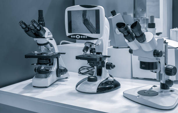
Choosing between SEM and optical microscopy isn't just about which one's more powerful—it's about matching the right tool to your specific analysis needs. While both reveal details invisible to the naked eye, they work in fundamentally different ways and excel in different situations. What Makes Optical Microscopy the Go-To Choice Optical microscopy remains the workhorse of many labs because it's straightforward, cost-effective, and surprisingly versatile. You're essentially using visible light and glass lenses to magnify samples, which sounds simple but works brilliantly for countless applications. Here's when optical microscopy really shines: Live sample observation: Want to watch cells divide or organisms move? Optical microscopes let you observe living specimens without killing them first Color differentiation: This is huge for pathology and materials science where color tells you everything about tissue types or material composition Quick turnaround: No special prep needed in many cases, just mount your sample and start looking Budget-friendly analysis: When you're processing dozens of samples daily, the lower operational costs add up The catch? You're limited to about 1,000x magnification, and you won't see much surface detail beyond that. Scanning electron microscopy brings the big guns when you need to dive deeper literally. Instead of light, you're firing electrons at your sample and reading how they bounce back. This completely changes what you can see. Professional SEM Lab Services become essential when your project demands: Extreme magnification: We're talking 100,000x or more, revealing nanoscale structures that optical microscopy can't touch Surface topology mapping: Those stunning 3D-like images you've seen of pollen grains or microchips? That's SEM showing every ridge, crater, and texture in incredible detail Material composition analysis: With the right detector setup, SEM tells you what elements are present and where they're located Failure analysis: Engineers rely on SEM to find microscopic cracks, corrosion patterns, or manufacturing defects in failed components The tradeoffs here are real, though. Sample prep takes longer, you can't examine living specimens, and the equipment costs substantially more to purchase and operate. Think of it this way: optical microscopy answers the "what" questions efficiently, while SEM tackles the "why" and "how" questions at a deeper level. Start with optical microscopy for general identification, counting, or measurement tasks. It's perfect for biological research, quality control in manufacturing, or educational settings where speed and accessibility matter. Move to SEM when you're investigating surface phenomena, need compositional data, or when your optical microscope hits its resolution limit and you're left squinting at blur. Industries like semiconductor manufacturing, forensic science, and advanced materials research couldn't function without it. The reality is that many labs use both, letting each technique do what it does best. Your specific application not just the magnification needed, should drive your decision.When SEM Takes the Lead
Making the Right Call for Your Project


Want to add a comment?